Microplastic Microcosm
March 11, 2024
It’s no secret that plastics and microplastics (MP) have spread like an unstoppable wave across our oceans. Marine plastics can be found covering the ocean’s surface and hiding in its deepest trenches. Their interactions with the larger species found in our ecosystems have been a topic of major concern. But, we must also focus on the smaller parts of our biosphere, namely the microbes and their interactions with plastics.
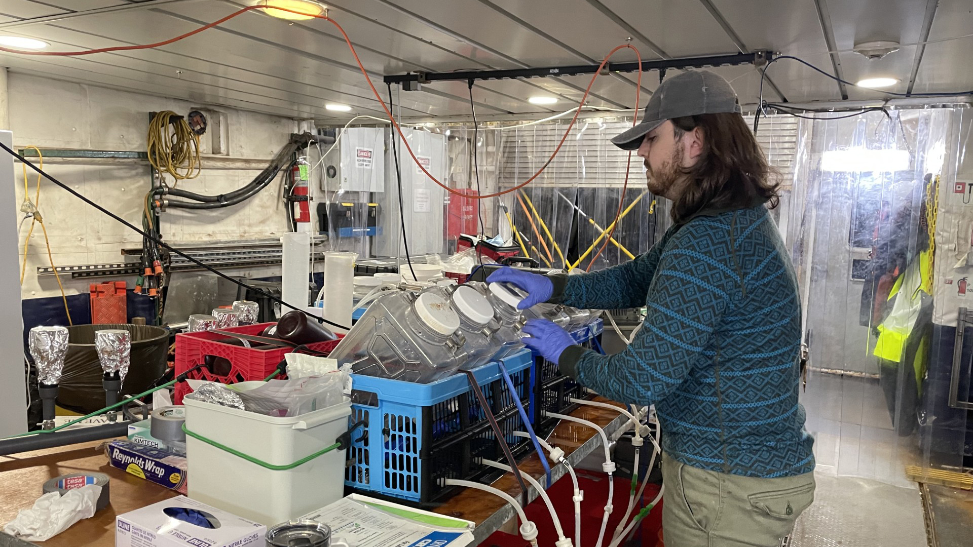
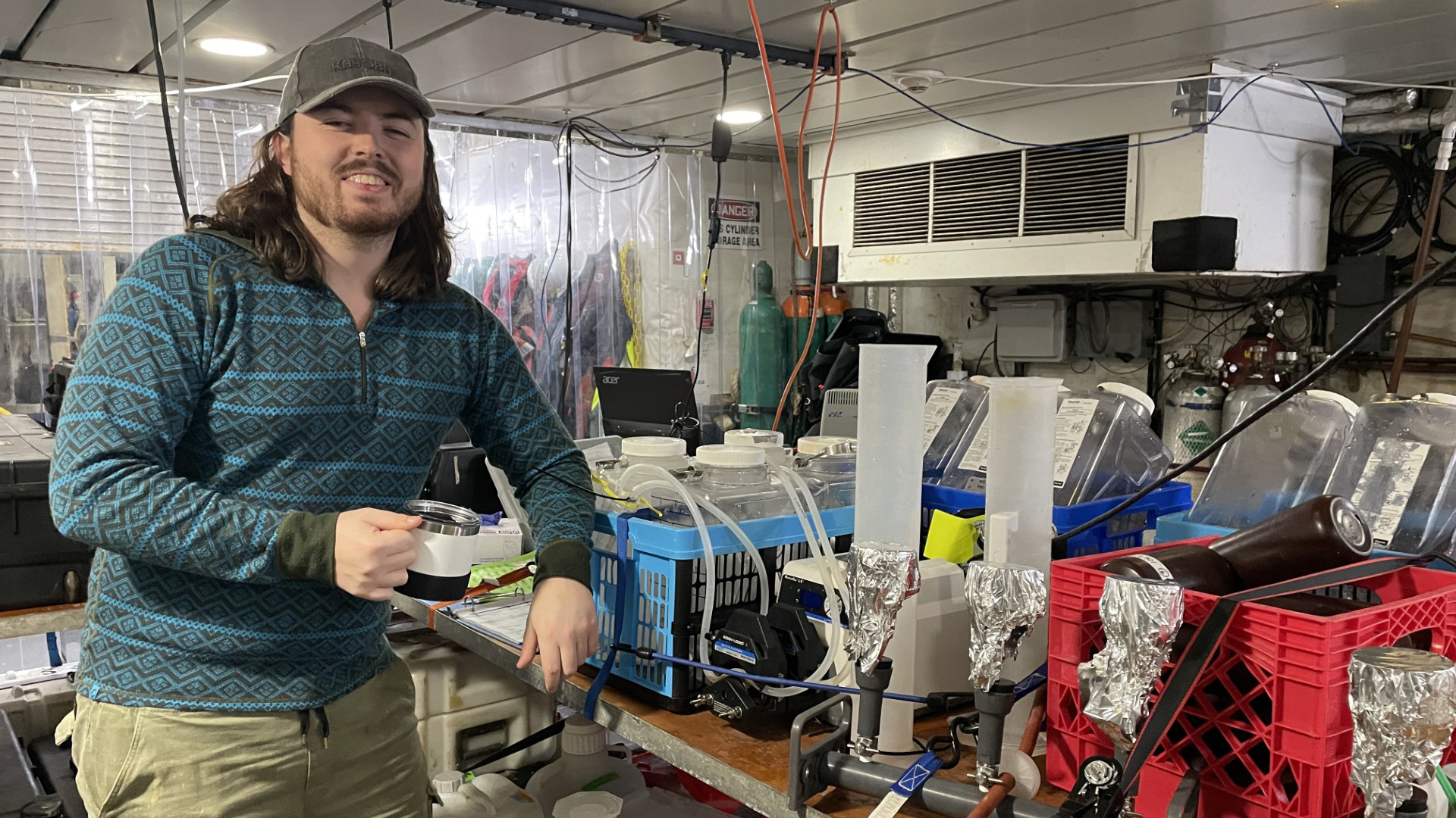
The “plastisphere” is a new niche for microbes to colonize. Not a surface made of natural material like rocks and leaves, but something man-made that offers new opportunities and potential hazards for microbes. In the arms race we know as microbial community development, the MP biofilms may select for the bacteria that are most resistant toward antimicrobial agents. Knowing about these antibiotic resistant genes and their presence on MP is key to understanding the eco-evolutionary impact of MP pollution. However, more research is needed to identify this new problem. In collaboration between the Marmib project and NOAA, I have been performing a microcosm experiment during the GO-SHIP A13.5 cruise down the African coast. Data from this project will be used to investigate the linkage between different types of MP, their microbial communities, and the presence of antibiotic resistant genes in plastic colonizing microbes.
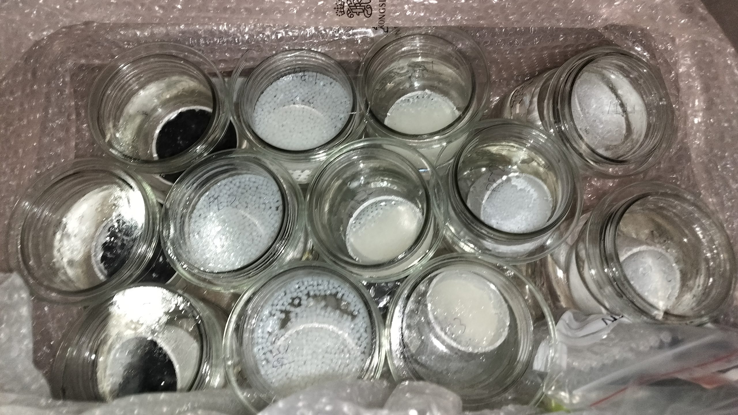
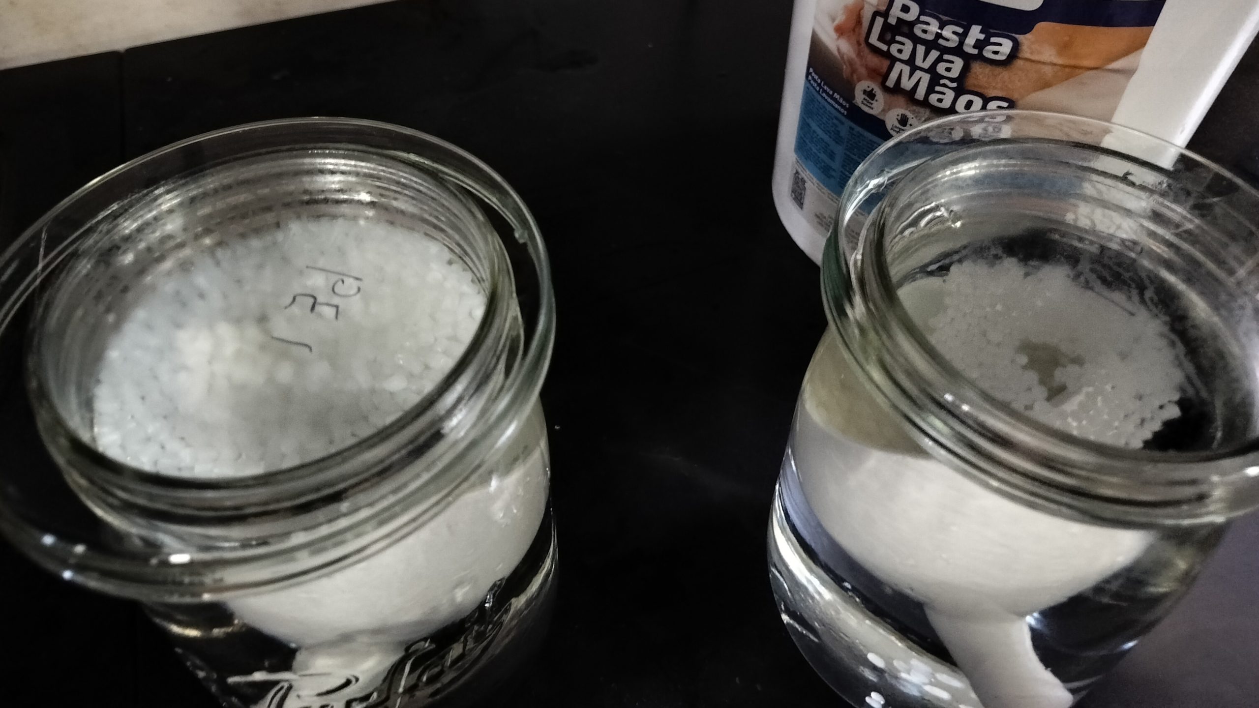
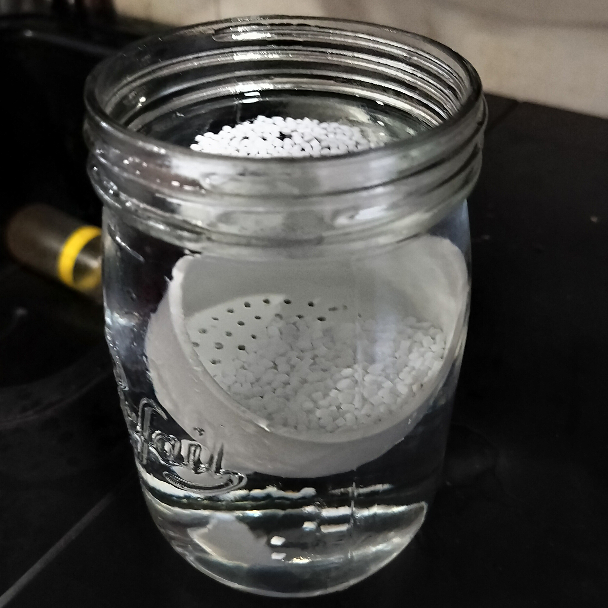
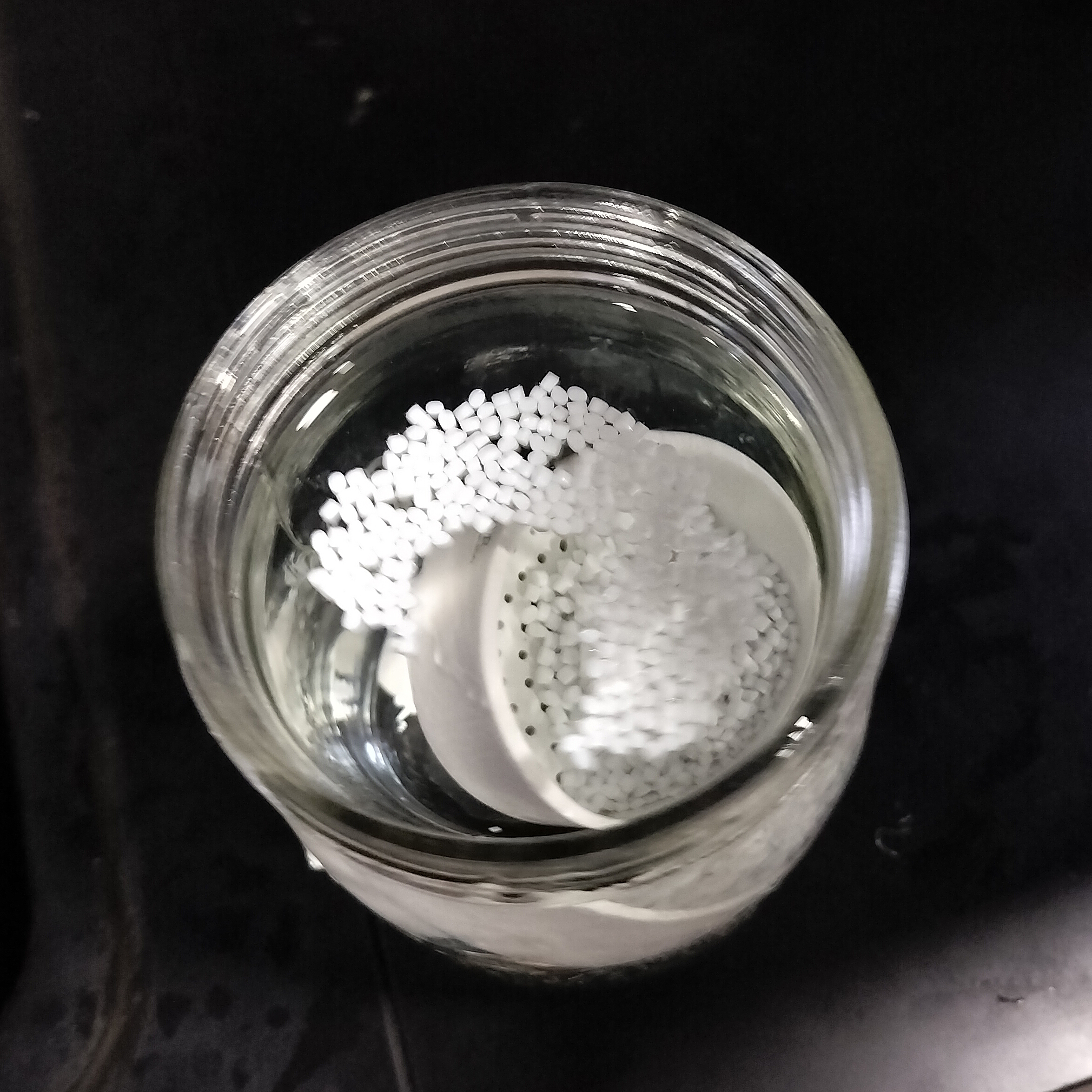
The microcosm experiment was set up in the form of 12 jars. The jars were split up into 4 groups of PE (polyethylene, the most commonly used plastic), PLA (polylactid acid, used for plastic films and in 3d printing), PET (polyethylene terephthalate, mainly used in bottle production) and rocks (control). Each jar was filled with grains of substrates corresponding to its group (that is, either one of the plastics mentioned above or rocks). The images below show the process described.
On the 7th of February 2024, the jars were filled with water from the ship’s underway system. Over the next month the water was exchanged once a week, using a funnel to make sure none of the substrate was lost. Each jar was covered by a petri dish to isolate it and prevent contamination. At the end of this experiment the samples were collected in different falcon tubes (as shown in the picture below) and frozen at -20 degrees Celsius, to be collected by one of Marmibs partners, Prof. Carlos Benzuidenhout (Director of the Unit for Environmental Sciences and Management at North-West University, South Africa), for DNA extraction and analysis.
The information we gain from this experiment will tell us how the microbial communities of the substrates differ and if there are any antibiotic resistant genes present.
Kristian Furnes, Biological Analyst, March 11, 2024
Master’s Student, University of Oslo, Norway
All pictures by Kristian Furnes unless otherwise noted.

