Say Hello to My Little Friend(s)
Signs of life from the abyssal zone
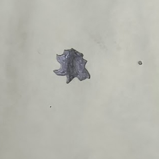
Every now and then, chemical oceanographers are confronted with very real evidence that there is, in fact, life in their water samples. Often called “bugs” in laboratory settings, their presence is generally ignored when gathering and analyzing samples. Sometimes, however, we accidentally capture them! This happened last Sunday, Feb 25th. My oxygen flask had two little visitors from the deep. Yes, I realize there are probably multitudes in most samples, but I could SEE these two. Remember Horton, the elephant who could hear the Whos? It’s an apt analogy to my excitement. I thought I had dark box-beetles or something. I didn’t know, I just knew they were alive and neat!
Of course, by the time I saw my little friends, I had already added the reagents necessary to run my experiment. This is the way of the scientist. The only reason I noticed them was that these two organisms were still moving through the cloudy precipitate inside the flask. So, what could they be? What would still keep on chugging along, brought to the surface from a depth of 4920m (that’s nearly 3 miles deep) and dosed with manganous chloride and sodium iodide as described in my earlier blog?
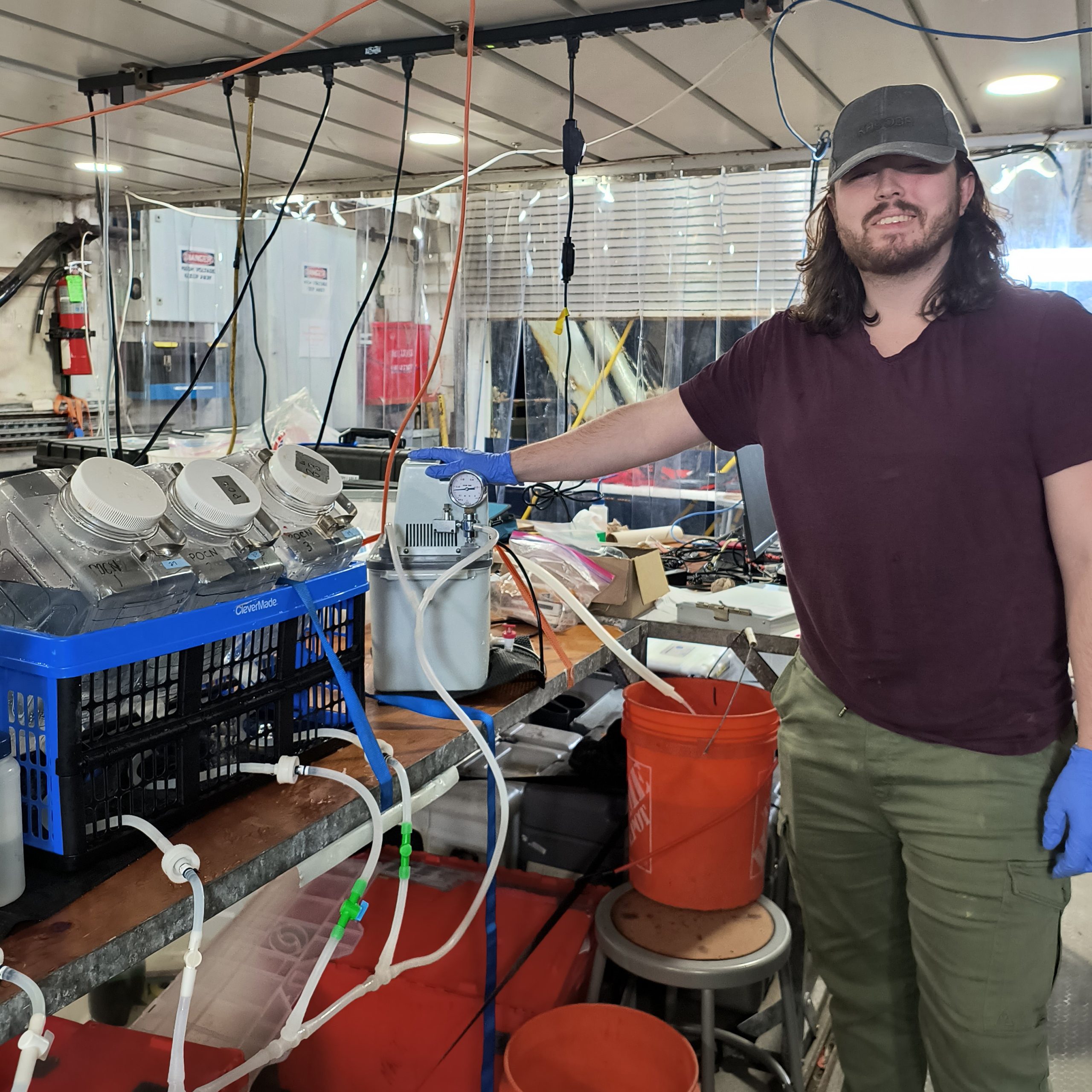
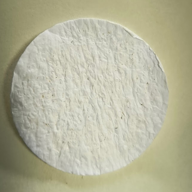
I had to defer to the internet, and others for possible answers. I asked everyone I could find awake if they knew what they were. I also requested help from biologist Kristian Furnes, masters student at the University of Oslo coordinating and making measurements of environmental DNA and other biological parameters on the cruise, to filter the remaining water collected at that depth.
Unfortunately, we could not find a microscope on board to examine the filter, but I did take a photo of the filter under a magnifying glass I found in the electrician’s workshop and there are many little specks and other items that are hard to determine what they are. When I return home, I plan to look at the filter under a microscope in case I can detect any additional organisms.
When I began my internet research, my initial search was looking for zooplankton which could exist at 4900m and be box-like swimmers. It suggested the following creatures: copepods, ostracods, amphipods, jellyfish, krill, or deep-sea larvae. Due to the depth, I mentally excluded phytoplankton, since they tend to be in the upper 100-200m of the water column. Since I was familiar with the suggested organisms, mostly long and shrimp-like, or gelatinous, I excluded most of them as well, but I did hold on to ostracods as a potential friend. Ostracods are round little swimmers.
Luckily someone with younger eyes, Eva Jundt, doctoral student from Texas A&M-Corpus Christi, told me it definitely wasn’t an ostracod. She could see more detail of the organism in the flask than I could and drew a picture of what she described as a “breadcrumb that swims.”
When I looked at Eva’s drawing of the original organism as she saw it, I remembered that there are other organisms beyond plants and animals. Foraminifera (or forams, for short) are amoeboid protists, single celled organisms that encase themselves in ‘shells’ of calcium carbonate and can exist in a huge variety of environments. When in the abyssal zone, they are generally considered to be benthic, living in or on the seabed. This sample was not from the true bottom of the ocean, but from around 610m above it. Geologists have used forams for years to try to find promising new sources of oil.
Forams, and their response to the lowering pH of the world’s oceans, was one of the ‘early indicators’ of the ocean acidification issue. Sadly, when I acidified my sample for analysis, my little friends disappeared forever. We could think about foraminifera as “canaries in the coalmine” due to in their inability to survive in acidic water. This same response occurs to many other organisms, and entire ecosystems. Snails, oysters, mussels and other sea creatures that build shells, even the corals in reefs, are highly affected by increasing acidification of our ocean. The increase of atmospheric CO2 entering our ocean from deforestation, cement processing, and the burning of fossil fuels contribute to the alarming level of ocean acidification we are detecting.
I still don’t know definitively what my little friends were, but I’ve accepted forams as my best answer. If you’d like to learn more about them, Wikipedia, the British Geological Survey, and many schools will have great information. I highly suggest watching an 8-minute YouTube video entitled, Foraminifera: Hard on The Outside, Squishy on the Inside.
Jennifer Aicher, O2 Team, March 5, 2024
University of Miami Rosenstiel School of Marine, Atmospheric, and Earth Science.

